
Sacral Hiatus - A Morphometric and Anatomical Study
Abstract:
To
accurate performance of epidural anaesthesia and analgesia it is important to
know the variations of sacral hiatus in dry bone. The present observational
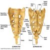
study conducted on one hundred dry human sacra to evaluate the anatomical and
morphometric variations of sacral hiatus. The most common shape of sacral
hiatus was inverted ‘U’ and ‘V’ respectively. Apex of sacral hiatus present at
3rd sacral segment in 78% sacra and base is at fifth sacral segment
in 89% sacra. The length of sacral hiatus is between 11-20 mm in majority
sacra, anteroposterior diameter is 4-6 mm in 63% sacra and transverse diameter
is 11-15 mm in of sacral hiatus. Anatomical variations in sacral hiatus can
leads to caudal epidural anaesthesia failure and procedure related
complications. Understanding these variations may improve success of caudal
epidural anaesthesia and decrease incidence of complications.
Keywords:
Sacral hiatus, caudal epidural anaesthesia, Morphometry.
References:
[1]. Kumar
V, Pandey SN, Bajpai RN, Jain PN,
Longia GS. Morphometric study of sacral
hiatus. J Anat Soc India. 1992; 41:7–13.
[2]. Lanier VS, Mcknight HE, Trotter
M. Caudal analgesia: An experimental and
anatomical study. American journal of Obstetrics
and Gynaecology 1944; 47(5): 633 – 641.
[3]. Letterman GS, Trotter M.
Variations of the male sacrum: Their significance
in caudal analgesia. Surg Gynecol Obstet.
1944; 78:551–5.
[4]. Manisha
B. Sinha, Mrithunjay Rathore, Human Prasad
Sinha: A Study of variation of sacral
hiatus in dry bone in central Indian
region; International J. of Healthcare and
Biomedical Research, Volume: 2, Issue: 4,
July 2014, Pages 46-52.
[5]. Nagar
SK. A study of sacral hiatus in
dry human sacra. Journal of Anatomical
Society of India 2004; 53(2): 18 -
21.
[6]. Standring
S, Newell RLM, Collins P, Healy JC.
In the back in; Gray’s Anatomy, The
anatomical basis of clinical practice. 40th
Edition. ISBN; 978-0-8089-2371-8. SPAIN, CHURCHILL
Livingstone Elsevier, 2008; pp; 724-5 2.
[7]. Standring
S, Ellis H, Healy JC, Johnson D.
Gray's anatomy. 39th ed. Vol. 1431. London:
Elsevier Churchill Livingstone; 2005. pp. 749–54.
[8]. Sekiguchi
M., Yabuki S., Satoh K., Kikuchi S.
(2004) an anatomic study of the sacral
hiatus: a basis for successful caudal
epidural block. Clin. J. Pain 20: 51–54.
[9]. Trotter M, Letterman GS, Gordon
S, Variations of the female Sacrum: their
significance in continuous caudal anesthesia.
Surg., Gynec. And Obst. 1944; 78:419-424.
[10]. Vinod kumar et al. Morphometrical
study of sacral hiatus. Journal of
Anatomical society of India 1992; 41(1):
7 – 13.
[11]. Waldman SD caudal epidural
nerve block; prone position in – Atlas
of interventional Pain Management, 2nd edn.
Philadelphia; Saunder 2004; 380-92.
